Differential Diagnosis of Heel Pain
Although heel pain occurs with a variety of injuries (e.g., calcaneal stress fractures and/or infracalcaneal bursitis), by far the most common cause of heel pain is plantar fasciitis. The word fascia is Latin for “band,” and the medial portion of the plantar fascia, which runs from the medial calcaneal condyle to the base of the hallux, represents the strongest and most frequently injured section of the band.
Until recently, it was assumed that excessive lowering of the medial arch in flat-footed individuals increased tension in the plantar fascia and overloaded the proximal insertion of the plantar fascia on the medial calcaneus. In fact, this increased tensile strain at this site was believed to be so great that it was thought to be responsible for the formation of a calcaneal heel spur. Although logical, recent research proves this is not the case, as a detailed histological study of 22 calcanei with heel spurs revealed that the bony exostosis forms at the origin of abductor digiti minimi and flexor digitorum brevis, not the plantar fascia.1
This research emphasizes the important clinical interactions that occur between the plantar fascia and the intrinsic muscles of the arch: The plantar fascia functions passively to store and return energy, while the intrinsic muscles play a more dynamic role in variable load sharing, working with the plantar fascia to prevent deflection of the arch during early stance and assisting with arch elevation during the latter portion of stance. This explains why the development of plantar fasciitis is not correlated with arch height and the best kinematic predictor of the development of plantar fasciitis is the speed in which the digits dorsiflex during the propulsive period.2
 Fig. 1. Flexor digitorum brevis home exercise.
The seated patient places an exercise band beneath the foot, traversing beneath the lesser toes up to the knee. Tension in the band is determined by the pulling force at the knee and the patient actively plantarfl exes the toes against resistance (arrow). To strengthen fl exor hallucis longus, this exercise is repeated beneath the big toe. To improve endurance, 8 sets of 40 repetitions are usually performed daily.When the flexor digitorum brevis is strong, it effectively decelerates dorsiflexion of the toes during the propulsive period while equally distributing pressure between the distal phalanxes and the metatarsal heads. Weakness of this small, but important muscle allows the digits to dorsiflex rapidly through larger ranges of motion, increasing the tensile strains placed on the plantar fascia. As a result, successful treatment requires decelerating the speed of digital dorsiflexion by strengthening not just the flexor digitorum brevis muscle, but also the flexor hallucis longus and flexor hallucis brevis. (Fig. 1) The speed in which the digits dorsiflex may also be lessened by shoe gear, such as Skechers or MBT, because the built-in rocker bottom present in these shoes limits the range and speed of digital dorsiflexion.
In addition to strengthening the digital flexors, chronic plantar fasciitis often responds well to low-dye taping and to custom and prefabricated orthotics (which are equally effective for the short-term treatment of plantar fasciitis).3 As demonstrated by Kogler, et al.,4-5 buttressing the medial longitudinal arch and incorporating rearfoot varus and/or forefoot valgus posts may significantly lessen tensile strains present in the plantar fascia.
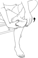 Fig. 2. Plantar fascia home stretch. This stretch is held for 10 seconds and repeated 30 times per day. The plantar fascia should be lightly massaged while performing this stretch.Other conservative treatment interventions include frequent stretching of the posterior calf musculature and the use of night braces. DiGiovanni, et al.,6 demonstrated that improved clinical outcomes occur with the simple addition of a home stretch. (Fig. 2) This stretch is held for 10 seconds and repeated 30 times per day.
Although deep-tissue massage may be helpful because it improves resiliency of the plantar fascia and may stimulate repair, care must be taken to avoid irritating the medial and lateral plantar nerves, which may be contused by overly aggressive cross-friction massage. When performed properly, deep-tissue massage coupled with stretches to restore first metatarsophalangeal joint dorsiflexion almost always results in a 10° increase in the range of hallux dorsiflexion. This is significant, since surgical release of the medial band of the plantar fascia has been shown to increase the range of first metatarsophalangeal joint dorsiflexion by 10°.7
Because of this, surgical release of the plantar fascia (which may result in a gradual destruction of the medial arch) should not be considered unless manual therapy fails to improve the range of first metatarsophalangeal joint dorsiflexion. The response to manual therapy can be evaluated with careful pre- and post-treatment measurements of hallux dorsiflexion. The efficacy of manual therapies for lessening plantar heel pain was proven in a randomized, controlled trial in which the addition of trigger-point massage to a conventional self-stretching protocol produced superior short-term outcomes compared to stretching alone.8
 Fig. 3. Baxter’s neuropathy test. When the nerve to the abductor digiti quinti is compressed, the patient is unable to abduct the fifth toe (A). MPN=medial plantar nerve; LPN=lateral plantar nerve; BN=Baxter’s nerve.Alternate causes of heel pain include enthesopathy from various autoimmune disorders, Baxter’s neuropathy, calcaneal stress fracture, and/or heel spur syndrome. The autoimmune disorders, such as rheumatoid and psoriatic arthritis, frequently produce pain and swelling at the plantar fascia origin, and are often misdiagnosed because the early signs are similar to those associated with mechanical plantar fasciitis.
Clinical clues suggesting autoimmune causes for heel pain are that these disorders tend to produce discomfort bilaterally, and the swelling tends to be more extreme. If psoriatic arthritis is the cause, skin plaques can often be seen on the hands or behind the ears. Suspected cases should be referred to a rheumatologist.
Another cause of heel pain is Baxter’s neuropathy. This condition represents a nerve entrapment syndrome in which the nerve to the abductor digiti quinti (also known as Baxter’s nerve) becomes inflamed beneath the proximal portion of the plantar fascia. Clinical signs of Baxter’s neuropathy include the reproduction of pain by abducting and dorsiflexing the forefoot for 30-60 seconds, a positive tourniquet test (i.e., pain is reproduced by inflating a blood pressure cuff placed around the lower leg to slightly above systolic pressure for 30 seconds) and/or the patient is unable to actively abduct the fifth toe on the involved side. (Fig. 3)
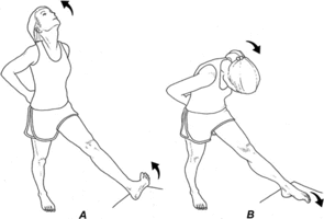 Fig. 4. Nerve glide test. To mobilize Baxter’s nerve, the patient places the heel on an elevated platform and then alternately extends the neck while dorsiflexing the ankle and toes (A), and then flexes the neck while plantarflexing the involved ankle and toes (B). Each cycle is performed for approximately 5 seconds and there should be minimal discomfort while performing this procedure.In addition to standard therapies to lessen inflammation, an alternate technique for treating Baxter’s neuropathy is to perform nerve glides on the nerve to the abductor digiti quinti. This is accomplished by heating the involved region, lightly massaging a 4-inch area directly over the site of entrapment (confirmed with Tinel’s sign), and then performing a series of light stretches in which the nerve is “flossed” back and forth in its tunnel. (Fig. 4) This technique has been proven to mobilize nerves in the upper extremity,9 and is believed to loosen adhesions responsible for maintaining the nerve in a fixed position.
If Baxter’s neuropathy is present, custom and prefabricated orthotics are often helpful since they may lessen the “scissoring” of the nerve between the long plantar ligaments and the plantar fascia. The exception to this is if an orthotic is made in which apex of the arch is placed beneath the sustentaculum tali. The proximally positioned arch apex may damage not just Baxter’s nerve, but also the medial and lateral plantar nerves. If an orthotic is to be used in the treatment of Baxter’s neuropathy, the laboratory must be instructed to place the apex of the arch beneath the medial cuneiform.
It is also possible that chronic heel pain is the result of an undiagnosed calcaneal stress fracture. A simple in-office test to rule out calcaneal fracture is the medial / lateral squeeze test. Because cortical bone in the calcaneus is so thin, medial and lateral compression of the calcaneus between the thumb and index finger produces significant discomfort when a stress fracture is present. To ensure accuracy, sensitivity to pressure should be compared bilaterally. If a calcaneal stress fracture does occur, it is important to identify the cause, such as underlying osteopenia / osteoporosis.
The final factor to consider in the differential diagnosis of plantar fasciitis is heel spur syndrome. The easiest way to differentially diagnose these two conditions is to ask the patient if they have increased pain while walking on the heel or the forefoot. Because plantar fasciitis is a propulsive period injury and heel spurs hurt during the contact period, patients with plantar fasciitis have more pain while standing on their toes, while patients with heel spur syndrome complain of pain when striking the ground on the involved heel. In fact, heel spur patients often make initial ground contact with the lateral forefoot in an attempt to lessen pressure beneath the heel during the contact period.
Because the treatment protocols for plantar fasciitis and heel spur syndrome are different, it is important to diagnose these two conditions correctly. Plantar fasciitis is treated with orthotics, stretches and exercises, while heel spur syndrome is treated with pocket accommodations, heel cups and well-fitting heel counters. Cortisone injections should be a last resort, especially in individuals with heel spur syndrome, because it may result in further degeneration of the calcaneal fat pad.
As with the majority of mechanical musculoskeletal conditions, treatment interventions emphasizing manual therapy, orthotics, stretches, and rehabilitative exercises almost always outperform popular, yet ineffective pharmacological interventions such as NSAIDs and corticosteroid injections.
Source: Dr. Thomas Michaud